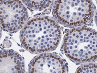
IHC for Ovarian follicle counts
Enumerating ovarian follicles is an effective way to estimate the extent of ovarian toxicity in female rodents exposed to xenobiotics. Differential follicle counts are useful in safety assessment bioassays and in interspecies extrapolation of ovarian toxicity. Counting the follicles in H&E-stained sections is labor intensive, tedious, and costly. Here, at TPA, we demonstrated that in rat formalin-fixed, paraffin-embedded ovary sections follicles of all degrees of maturity can be visualized by the use of antibody directed against proliferating cell nuclear antigen (PCNA). Follicles are easily distinguished from ovarian background with the ability to detect and identify primordial follicles being enhanced. This translates into a significant decrease in variability of follicle counts, labor, and cost.

PCNA-stained oocytes of primordial follicles in rat ovary (PCNA IHC). x160
IHC for Human in vitro air-liquid interface (ALI) models
ALI models create a fully differentiated, in vivo-like human bronchial epithelium. They provide a potential means to generate relevant data for evaluation cigarette smoke toxicity.

Proliferating cells in human ALI model (Ki67 IHC), x 160
Apoptotic bodies in human ALI model (Cleaved caspase-3 IHC), x160
Involucrin positive cells with signs of squamous differentiation in human ALI model (Involucrin IHC), x160
In Situ Hybridization
In situ hybridization (ISH) techniques allow detection of specific nucleic acid sequences in morphologically preserved cells or tissue sections. In combination with IHC, ISH can provide microscopic topological information about gene activity at the DNA, mRNA, and protein level.
Non-radioactive ISH for CYP1B1 mRNA, human brain. x40
Non-radioactive ISH for histone mRNA, mouse spleen. x100
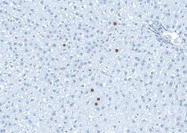
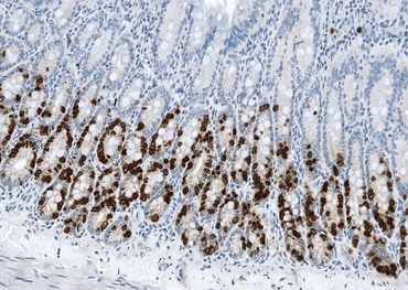
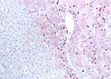

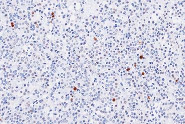
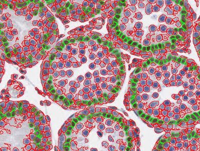
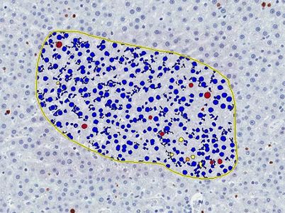
Image Analysis
IHC-stained sections are scanned and digital images are obtained by Aperio Scanscope System (Aperio Technologies, Inc., Vista, CA). In these images, intensity of staining, proportion of immunostained area and other parameters are evaluated with Positive Pixel Count Algorithm. This algorithm quantifies the amount of specific stain present in a digital image by evaluating average intensity of all pixels (Iavg); values of Iavg are then used to calculate optical density (OD). The Nuclear Algorithm evaluates numbers (%) of positively stained nuclei and quantifies the average staining intensity in individual cells.

Automatic counting of Sertoli cell nuclei stained with Sox9. Rat testis. x80
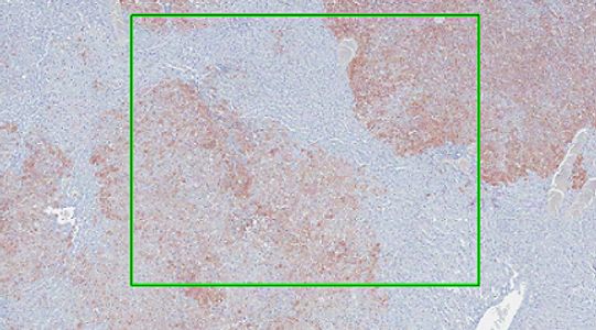
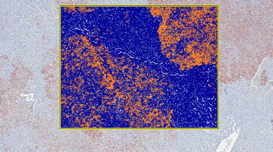
Automatic counting of proliferating cells in the outlined area of rat liver section (Ki67 IHC). x80
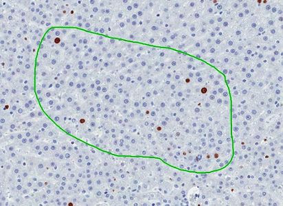
Automatic measurements of positively stained tissue and intensity of staining in the outlined area of GSTP-stained liver section. x4
Automatic counting of proliferating cells in human ALI model (Ki67 IHC). x160
Automatic measurement of positively stained area (fat droplets) in the mouse liver (osmium tetroxide staining), x40
IHC of Free-Floating Sections
When performing IHC on very thick tissue sections (~40µm) free-floating is the method of choice. This allows for better penetration of the antibodies and 3-D reconstruction of large structures such as axonal projections or vessels.

Each brain is sectioned into a 24 well plate. With each of the 24 wells containing sections representative of the entire brain. ~12-15 sections per well
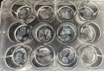
One well from the 24 well plate is sorted into 3 wells on in a 12 well plate for staining. ~4-6 sections per well.

After staining is complete the sections are placed onto slides using a paint brush. Each well corresponds to one slide.
After sections are placed onto slides fluorescence microscopy is used to image the tissues. Tissues are sometimes double stained as seen here with GFAP to label astrocytes and glial cells as well as DAPI to label all nuclei. Tissues can also be triple stained as seen here with DAPI (nuclei), NeuN (Neurons), and IBA1(microglia/macrophages).
Western Blot Analysis
This is an excellent technique for quantifying total amounts of specific proteins in any tissue. This technique is a useful alternative to IHC when the exact location of the protein is not needed, only the quantity.

Western blot of protein from hippocampus cell lysates, synaptophysin (green, 38kDa) and beta actin (red, 42kDa).